 |
Obrázok 1 Využitie
elektrokardiogramu v diagnostike kardiálnych aj nekardiálnych porúch Figure 1 The utilisation of the electrocardiogram in the diagnostics of cardiac and non-cardiac disorders |
| Obrázok 2 Dynamika zmien vlny
T, segmentu ST a vzniku kmitu Q pri akútnom infarkte myokardu. So súhlasom
vydavateľstva Lippincot Williams and Wilkins (13). Figure 2 Dynamics of changes of the T wave, ST segment and emergence of the Q oscillation in acute myocardial infarction. With permission of Lippincot Williams and Wilkins (13) |
|
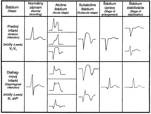 |
Obrázok 3 Vývoj
elektrokardiogramu pri akútnom infarkte myokardu – elektrokardiografické štádiá
akútneho infarktu myokardu Figure 3 Development of the electrocardiogram during acute myocardial infarction – electrocardiographic stages of acute myocardial infarction |
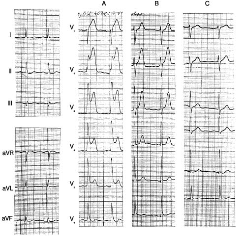
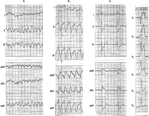
| Obrázok 4 Včasný
elektrokardiogram pri akútnom infarkte myokardu Figure 4 Early electrocardiogram during acute myocardial infarction |
|
 |
Obrázok 5 Elektrokardiogram
59-ročného pacienta s vazospastickou angínou pektoris Figure 5 Electrocardiogram of a 59-year old patient with vasospastic angina pectoris. |
 |
Obrázok 6 Elektrokardiogram
57-ročného pacienta so syndrómom včasnej repolarizácie. V končatinových
zvodoch prítomné dva komplexy s obrazom W-P-W s rôznym stupňom preexcitácie Figure 6 Electrocardiogram of a 57-year old patient with the early repolarisation syndrome. In the unipolar leads two complexes with a W-P-W morphology with a different degree of pre-excitation are present. |
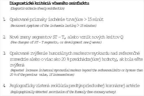 |
Obrázok 7 Diagnostické
kritériá veasného reinfarktu Figure 7 Diagnostic criteria of early reinfarction |
 |
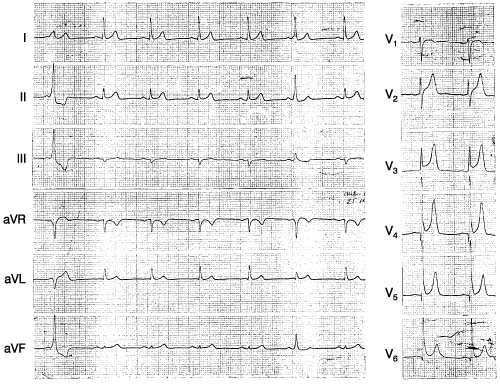
Obrázok 8 Elektrokardiogram
62-ročného pacienta. Figure 8 Electrocardiogram of a 62-year old patient. |
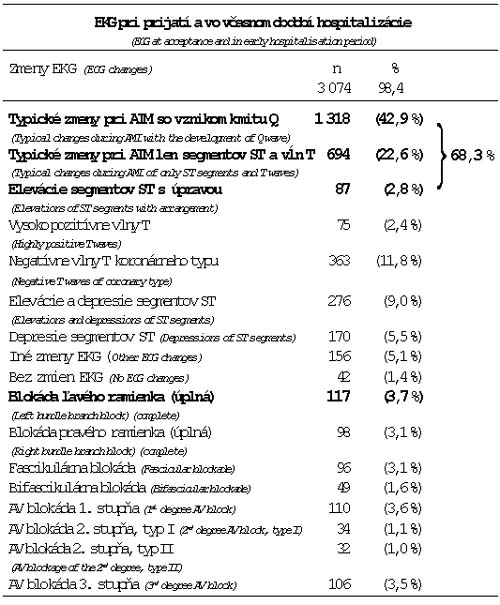 |
Tabuľka 1 EKG pri prijatí
a vo včasnom období hospitalizácie (doteraz nepublikované údaje) AIM – akútny
infarkt myokardu Table 1 ECG at the admission and in the early stage of hospitalization (unpublished data) AMI – acute myocardial infarction |
| Tabuľka 2 Korelácia
elektrokardiografickej a angiografickej lokalizácie. Table 2 Correlation of the electrocardiographic and angiographic localisation. |