 |
Obrázok
1 Klasifikačné systémy pri aortálnej disekcii |
 |
Obrázok
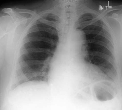
2A Skiagram hrudníka z 11. novembra 1999. Srdcový tieň je obojstranne rozšírený,
s aortálnou konfiguráciou, KTI = 0,57. Pľúcny parenchým je bez ložiskových zmien,
bilaterálne sú výraznejšie hily a tiež bronchovaskulárna kresba v dolných
pľúcnych poliach. |
 |
Obrázok
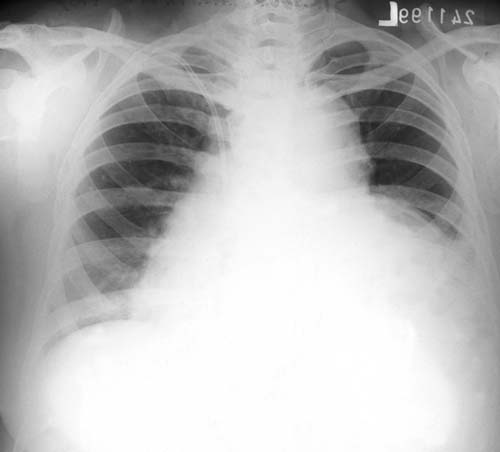
2B Zreteľné progresívne rozšírenie srdcového tieňa, pričom ľavá hranica
siaha až po laterálnu stenu hrudníka (24. novembra 1999). KTI = 0,75 |
 |
Obrázok
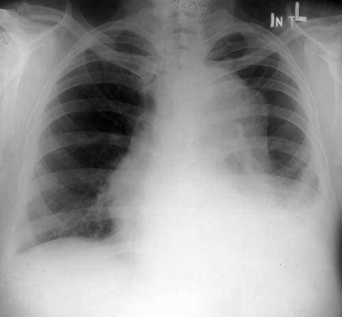
2C Výrazne rozšírený tieň hrudnej descendentnej aorty na skiagrame hrudníka zo
17. decembra 1999. |
 |
Obrázok
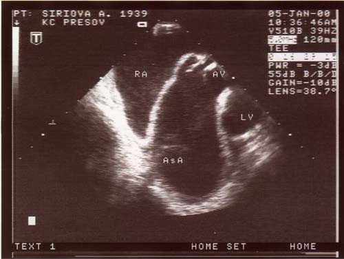
3A Dilatovaná ascendentná aorta pri transezofageálnom vyšetrení
v transverzálnej rovine bez príznakov disekcie. Aortálna chlopňa je
kalcifikovaná, so zníženou separáciou a maximálnym gradientom PG = 15,5 mHg
a Vmax = 1,97 m/s. |
 |
Obrázok
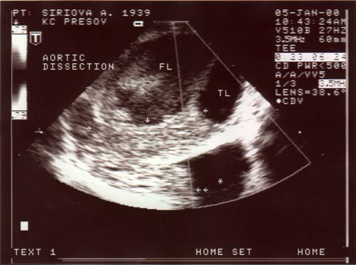
3B V distálnej časti hrudnej descendentnej aorty je zobrazený skutočný
kanál (TL) a trombus (ohraničený jednoduchými šípkami) v disekčnom
kanáli. V paraaortálnom priestore (*) je tiež zachytená trombotická masa
(dvojitá šípka), ktorá je v kontakte so zadnou aortálnou stenou. |
Obrázok
4 Brušná aorta s priemerom 3,2 cm. Disekčná membrána jednoznačne
vizualizovaná nebola, avšak farebným (i pulzným) dopplerom bol oddiferencovaný
dorzálne uložený skutočný kanál (TL), široký 1,5 cm, znázornený v systole
farebným tokom. Disekčný kanál (FL) je bez prítomnosti krvného toku. |
|
 |
Obrázok
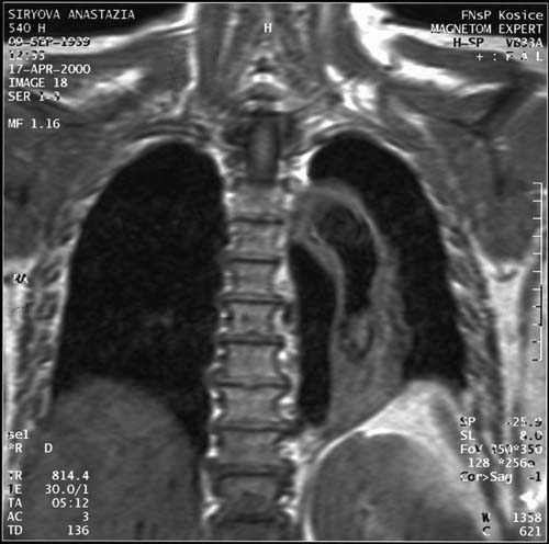
5 Hrudník vo frontálnej rovine pri vyšetrení nukleárnou magnetickou rezonanciou.
Zachytená je dilatovaná hrudná aorta od odstupu a. subclavia l. sin. až po bránicu.
V centrálnej časti je zachytený skutočný kanál; v dolnej časti vo
falošnom kanáli sa nachádza trombus. |
 |
Obrázok
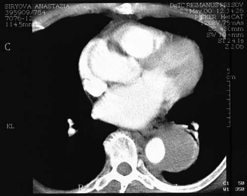
6 Špirálový CT sken hrudníka s kontrastom. Transverzálny sken zachytáva
dilatovanú, kontrastom vyplnenú ascendentnú aortu a dilatovanú descendentnú
aortu, v ktorej je pravý lúmen vyplnený kontrastom; falošný kanál je menej
denzný. Dorzálne je zachytená komunikácia s intrapleurálnym priestorom. |
Tabuľka
1 Výsledky vyšetrenia punktátu z perikardiálneho vaku |
|
Tabuľka
2 Výsledky markerov na onkologické ochorenia |
|
Tabuľka
3 Percentuálny výskyt menej typických a atypických prejavov disekcie
aneuryzmy aorty |
|
Tabuľka
4 Senzitivita a špecifita zobrazovacích metód pri disekcii aorty |